About
GAMMA CAMERA
The gamma camera, also called scintillation camera, is the most commonly used imaging device in nuclear medicine. It simultaneously detects radiation from the area of interest / organ and enables the acquisition of dynamic as well as static images of the area of interest in the human body.
Various scans done by gamma camera:
- Stress tests – MPI / stress thallium
- Myocardial viability scan
- Bone scans
- Thyroid scans
- Testicular scan
- Gallium scan
- Renal scans / DTPA
- DMSA scan
- HIDA scan
- Lung scan
- Lymphoscintigraphy
- Positron emission tomography
- Salivary gland imaging
- Infection imaging
- Parathyroid sestamibi imaging
.png)
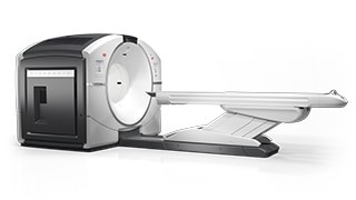
PET CT
A positron emission tomography (PET) scan is a nuclear imaging test that uses radioactive material to determine the metabolic and biochemical functioning of the tissues. The radioactive material is injected into the vein, and areas with abnormal metabolic activity will show increased uptake of the material, thus indicating the possibility of a disease. Changes in the biochemical properties can help identify the disease earlier before any structural or anatomic changes have happened in the tissues, which appear only later in an imaging process.
A. MALIGNANCIES
- Endocrine malignancies
- Muscular skeletal oncology
- Carcinoma lung
- Brain tumors
- Head and neck malignancies
- Carcinoma breast
- Hepatobilliary malignancies
- Gastrointestinal malignancies
- Gynaecological malignancies
- Genitourinary malignancies
B. NEUROLOGY
- Dementia imaging
- Epilepsy imaging
C. CARDIOLOGY
- Myocardial viability
- Myocardial infection imaging
D. PYREXIA OF UNKNOWN ORIGIN
HIGH DOSE THERAPY WARD
- Radio-iodine therapy
- PSMA therapy for prostate for cancer
- DOTA therapy for neuroendocrine malignancies